Early diagnosis of glaucoma, fundamental

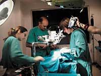
The prestigious American Glaucoma Foundation has awarded the project of the Faculty of Medicine of the UPV. This organization has awarded for the first time a European research.
The objective of the project is to create animal models with glaucoma in which the cellular and molecular mechanisms causing the disease can be investigated. This group works with pigs.
They aim to define methodologies to prevent the disease or at least allow a rapid diagnosis and develop therapies to curb the blindness caused by the disease. Thanks to the award, researchers will receive a total of $120,000 in two years.
The team led by Elena Barrio, which has eight researchers, has recently received the first prize of the ONCE in the 3rd edition of the International Awards for Biomedicine, R&D and New Technologies for the Blind.
Glaucoma
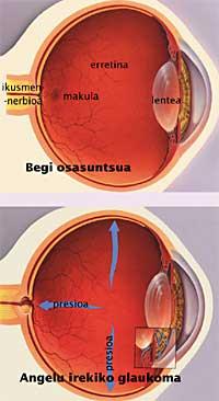
The disease suffered by the eyeball is characterized by irreversible damage to the fibers of the optic nerve. This is due to increased internal pressure of the eye.
Glaukoma comes from the Greek and refers to the green color that the pupil takes (glaukos = verdel).

But why is glaucoma formed? The structure of the eyeball feeds humor inside the eye. Humor is a totally transparent or fluid liquid that is pierced by light. Thus, light affects the retina without interference. Liquid humor forms in the ciliary body and moves through the pupil to the front chamber of the eye. There it feeds the anterior surface of the lens and the cornea. It is a very simple traffic. But when it gets upset, serious problems can arise.
If more fluid enters the front chamber of the eye than it can come out, the pressure increases and this pressure is supported by the fibers of the optic nerve. The pressure of intraocular humor varies from person to person, usually between 12 and 21 mmHg.
Glaucoma is one of the most common diseases in the eye; every year two million new cases are diagnosed in the world. Family history, age, and race are some of the determinants of the disease. In most cases of glaucoma, eye pressure increases causing the death of retinal cells until the patient becomes blind.
In addition, specialists who treat glaucoma face a serious problem: no symptoms appear until the disease is very advanced and, at the moment, it is not known why cells die.
The sooner glaucoma is detected, the more effective the possibility of treatment is. However, in diseases that initially have no symptoms, the only way to diagnose them is through an ophthalmologist's examination.
Therefore, taking into account that early detection is essential to prevent vision loss, a periodic visual review should be performed. Also, once a certain age is reached, about 50 or 60 years, we should all analyze the view with a certain frequency (measure the internal pressure of the eye, etc. ), because the same age is a risk factor.
Types of glaucoma

Although ophthalmologists distinguish dozens of types, the most basic are only three:
- Congenital glaucoma: passes from parents to children. Immediately after birth, the child, in addition to emitting numerous tears and photophobia, has a large eyeball.
- Acute or angle-closure glaucoma: It appears abruptly and produces a lot of pain as if it were stuck in the eye. Vision decreases sharply, the patient sees it blurred.
Chronic open-angle glaucoma: it is the most frequent and causes failures in the fluid removal system. Gradually and without symptoms, early detection is not easy. It can only be diagnosed by measuring the internal pressure of the eye.
The symptoms of congenital glaucoma and acute glaucoma are well known since the onset of the disease, unlike chronic glaucoma. In the latter, at first no symptoms are perceived and when they appear, as the optic nerve is usually damaged, the sight is lost: only the things that are in front are seen well, while the sides or those that look through the contour of the eye are hardly visible. This loss of vision is becoming more severe if there is no solution.


