
Clear brain tumors
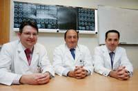
Malignant brain gliomas often have a poor prognosis, although they are used in combination with surgery, radiation therapy, and chemotherapy. In fact, the total removal of the tumor is often not achieved, which is very important to cure the disease. According to Dr. Ricardo Díez Valle, a specialist in the Department of Neurosurgery at the University Clinic of Navarra, experience shows that in patients who have achieved total removal of the tumor there are greater possibilities of removal of remains of tumor cells stopped by chemotherapy and radiation therapy.
One of the main problems for the extraction of gliomas is the difficulty in separating the tumor from healthy brain tissue, that is, knowing where to cut it. But a group of German neurosurgeons led by Walter Stummer developed a new technique that solves this problem. According to the work published by this group in the scientific journal Lancet Oncology, the use of an operating microscope with a fluorescent light module allows obtaining a complete extraction of malignant brain tumors in 67% of cases. This paper states that with conventional surgical techniques total removal is achieved in 30% of cases and that, after six months, 41% of patients treated with the new technique remain without trace of the tumor, while with conventional techniques it achieves 20%.
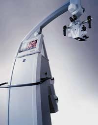
Well, the University Clinic of Navarra is one of the first hospitals in the State to obtain a microscope of this type. The state-of-the-art Pentero microscope is the highest equipment in surgical, optical and imaging technology.
According to Miguel Manrique, director of the Department of Neurosurgery of the University Clinic of Navarra, the new microscope serves for the extraction of all types of brain tumors and cerebral vascular malformations, as well as for epilepsy surgery in relation to mental injuries.
The new microscope also offers important advantages for the intervention of cervical arthrodesis and cervical, dorsal and lumbar disc hernias. In fact, "it offers greater speed and clarity, reducing wounds and the intervention is milder for the patient," says Dr. Manrique.
Separating limits
According to Dr. Díez Valle, the difficulty of extracting brain gliomas with conventional methods is especially at the edges of the tumor, as it is in contact or embedded with healthy brain tissue. And with conventional surgical views or microscopes it is very difficult to separate the tumor from the rest of the healthy tissue. With the new equipment, and thanks to the technique developed by the team of Stummer, the surgeon can easily separate the tumor from the brain tissue, completely removing the tumor, without causing brain damage, in place and size.
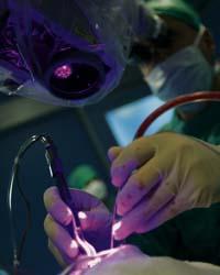
Tumor boundaries are separated by oral administration of a contrast, 5 aminolebulin acid. This substance is a modified amino acid that allows when illuminated with fluorescent light to appear in pink the cells in reproductive phase, that is, the tumor cells, and in blue the remaining. This substance has no side effects and is not marketed in Spain for this use, but can be obtained with the authorization of the Ministry of Health.
Thanks to the fluorescence module that incorporates the microscope, it is enough to press a button at any time to move from normal white light to a fluorescent vision of the brain. The surgeon can remove what he sees in pink without touching the healthy blue brain tissue.
In the operating room, neurosurgeons have the support of an initial navigation system to locate the tumor and know exactly where the brain from which the intervention should begin. The browser provides a map of the patient's brain thanks to information obtained in an MRI before the operation.
The equipment uses bio-based technology. In fact, it distinguishes cells according to their reproductive status or absence. The contrast substance marks the cells in reproduction. They have confirmed this in the department of Pathological Anatomy of the University Clinic of Navarra. Samples of tumors extracted during interventions have been analyzed there and confirmed to be tumor cells at the border with healthy brain tissue.
Therefore, they consider that the fluorescent light technique is the most suitable for this application and that it will contribute to significantly improve the prognosis of patients with malignant gliomas. "Thanks to the new technique we have managed to perform a complete removal of tumors that we would have previously considered almost impregnable," explains Dr. Díez Valle.
Zu idazle
Zientzia aldizkaria