

Looking inward
X-rays at the base
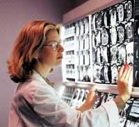
XIX. At the end of the 20th century, German physicist Wilhelm Conrad Röentgen discovered a major impact on medicine: X-rays. When he found them he didn't know what they were, so he called them X.
X-rays are electromagnetic waves capable of crossing different types of materials. Humans cannot see them at first glance, but being able to darken photographic emulsions, it was quickly used in medicine to perform X-rays.
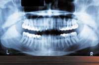
X-rays are images obtained as a result of X-ray action. X-rays pass through the body and reach specially treated plates. These plates are similar to those of the photos and when touched by x-rays a negative photo is taken.
X-rays easily pass through the body's soft tissues: blood, skin, fat, and muscles, and the plate looks dark gray. The bones or tumors, on the other hand, are more compact and being crossed by fewer X-rays, appear on the blank plate. For example, when the bone breaks, the x-ray beam crosses the broken area and looks like there is a black line on the white bone on the plate.
X-rays can be simple or with contrast. That is, the part of the body that needs to be looked through X-rays is sometimes not opaque enough, so products that cause contrast are introduced into the body by the mouth and veins.
Tomography: Computer X-ray
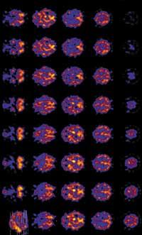
Another X-ray application is tomography, a technique that combines x-ray technology and computers. When common x-rays are performed only shadows are seen, for example, the tumor inside the body can be detected by x-rays, but it is not known to what depth it is. The doctor can then perform a new x-ray and have a shade obtained from another point of view. This can determine the approximate depth of the tumor. It can be done from many angles and the doctor can determine the tumor's position and shape. However, the doctor has limitations because he or she cannot process all x-rays at once. On the contrary, the computer can convert the two-dimensional images obtained by X-ray into three-dimensional, obtaining the exact depth and size of the tumor.
Scanners are used for tomography. The patient is placed in the center of the scanner, usually lying on a bed table. When the scanner starts to turn, at very short moments small doses of x-rays are passed from the body. The tissues of the body absorb these x-rays, the scanner detects them and sends information to the computer. This converts the information into an image for further analysis by the radiologist.
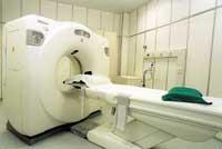
In the images of the scanners the body is seen as a filleting. Since the x-rays that are directed to obtain images of tissues or organs begin to rotate around the tissue or organ, images and details taken from different points of view can be obtained. Scanners provide accurate images of any part of the body, whether bones, muscles, fat, or organs. X-ray information is collected on a computer that performs two-dimensional and three-dimensional projections. The scanner is much more accurate than normal X-rays and although in a single session many images are collected, the patient receives fewer radiations.
In fact, in scanners the x-ray beam revolves around the body. As with simple x-rays, contrast techniques can be used in tomography. The scanners are performed mainly for the diagnosis of tumors, the study of hemorrhages and the detection of injuries or damage.
Wave diagnosis
Another observation system within the body is that of magnetic resonances. In magnetic resonances, the simultaneous help of large images, radio frequencies and computers allows accurate images of the inside of the body.
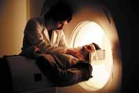
Resonance machines are usually large, tubular, capable of generating strong magnetic fields around the patient. These devices have, on the one hand, an electromagnet that produces a static magnetic field and, on the other, a radio frequency coil that generates a rotating magnetic field. Therefore, a powerful magnetic field is created around the patient when starting work. At the same time, radio waves are directed to the patient from a scanner. These waves ring the nucleus of the body's hydrogen atoms and change from their normal position. The cores send the radio signal as they return to their normal position. The computer collects, analyzes and transforms these signals into two-dimensional images. The image appears on the screen.
When working with strong magnetic fields, patients should remove all metal objects. Medications are also sometimes used to induce contrast through the veins of the arms or mouth. Once the patient is lying on a table, they introduce it into the tubular tool. During the observation process the patient remains alone in the room, but can talk to the doctors and is treated at all times behind the glass. When the scanning process starts, as the magnetic field is created and the scanner starts sending radio waves, large noises are heard inside the tube. Therefore, in many cases, patients have headphones to reduce the sensation of noise and listen to the orders of technicians.
Magnetic resonances are common to see the state of the heart, brain, liver, pancreas, male and female reproductive organs, and other soft tissues. It is also a suitable system for detecting blood flow, detecting tumors and numerous types of cancer, and detecting bone lesions.
The first resonance images were obtained in 1977. Despite initial doubts, experience shows that nuclear magnetic resonances do not harm man.
Beneficial radiation
Nuclear medicine is a specific and differentiated field of radiology. It uses radioactive substances or radiopharmaceuticals to know the state of the tissues. Images of nuclear medicine are the result of a multidisciplinary mix: chemistry, physics, mathematics, computer technology and medicine. This radiology area is used to study details in the early stages of disease.
X-rays go through soft tissues, such as intestines, muscles, or blood vessels, so substances are used to get contrast in nuclear images. Nuclear images analyze the structure and function of organs.
Nuclear medicine scanners are performed to study many body tissues and organs. Depending on the type of scanner different technologies, radiopharmaceuticals and techniques can be used. First the radiopharmaceutical is administered, then the images are taken and finally the interpretation of the images is performed. The process seems simple, but, depending on the case, from the administration of the radiopharmaceutical to the imaging can take a few minutes or a few days. The time to obtain the images may also be like this, sometimes it may vary a few minutes, but others may require hours.
The heart scanner is one of the most frequent scans through nuclear medicine. The control of the electrical activity of the heart is done by placing electrodes to the patient. Radiopharmaceutical is given through the veins. This radiopharmaceuticals blood cells, so passing through the heart can be seen by a scanner. The Gamma camera, a device used to scan patients with low doses of radioactive materials, gets images of the heart. When imaging, the camera measures the amount of radioactive substance that absorbs the heart. Once all the images have been obtained, the radiopharmaceutical instruments are removed from the veins and the process is completed. In most cases, these scans are done with the patient in a quiet position, but the doctor may need images of the time the heart is working intensively, so he is encouraged to perform physical exercises while monitored.
Ultrasound in ultrasound
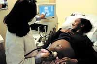
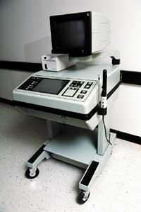
One of the most used techniques to see what is happening inside us are ultrasound. Ultrasound scans use high-frequency sound waves (ultrasound) and a computer to make images of tissues, blood vessels, and organs. Due to the use of ultrasound, the terms ultrasound and sonography are used to designate this technique.
Ultrasounds are done by wetting the outline of the body that needs to be looked at first with a gelatinous substance. This gel acts as a conductor. A transducer is used for sending ultrasound waves. The sound of the transducer is reflected in the structures inside the body and a computer analyzes the information of those sounds and creates an image on the screen.
Ultrasounds are usually performed for observation of the organs and blood vessels inside. The belly, breasts, pelvis, prostate, scrotum, thyroid, etc. are usually performed to observe the vascular system. During pregnancy it is common to perform ultrasound scans to see the development of the fetus. In fact, ultrasounds are a safe indoor observation system, which does not seem to harm ultrasound.
Interior
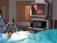
More aggressive techniques are also used than previous ones: endoscopic techniques. They are more crude because a certain invasion occurs in the body, but thanks to that invasion can become more effective, without causing excessive damage. These techniques include tubes or probes with a video camera inside the body. These cameras provide excellent images of the interior. Images can sometimes be seen on a screen or, in many others, from the other side of the tube, you can see the body directly.
Endoscopic techniques are often used not only for imaging and diagnosis, but also for surgical interventions. That's why these techniques can be so effective. Very small wounds. These probes introduce pipes, probes, catheters, etc. and the necessary operations are carried out. Many of the long and large seasonal operations that left great scars for life, today are performed by endoscopy. The main challenge of this type of surgery is therefore to find the most appropriate way to move catheters and probes within the body.
The operations performed in the veins, or those performed using the veins as a medium, are the best demonstration of endoscopic techniques. With a few millimetre catheter tucked into the veins, surgeons are able to reach places they just couldn't imagine, such as entering the aorta at the waist and reaching the heart. Depending on what they carry at the end of the tube or catheter, they can do one or another task: place valves in the veins, make some incision, send images by ultrasound… As you can see, as the technology increases the possibilities of small-scale work, the possibilities and resources of medicine are also far ahead.
From the patient perspective The advancement of technology has not only benefited doctors, but also patients. Diagnostic tests are getting faster, more accurate and, most importantly, less harsh than ever before. As indicated in the following lines, this is evident. However, test attendance remains harsh, not only because of the severity that can occur until the diagnosis is known, but tests are not usually very sweet. Simple assistance can be a source of nerves for many people, such as the need to be fasting and the coldness of the product entering your veins. In some cases it is very uncomfortable to take the necessary purge or fasten it. For the elderly or simply very nervous, there are times when you have to keep your breath, be still, ingest liquids of bad taste or, worse, overwhelm the tube that enters your throat and withstand the obstacle of the tube from the nose to the belly. What about the anguish caused by the resonances of the whole body? They do not cause harm, but many who have done so claim that they have never felt the feeling of being in the coffin closely. Therefore, technology is on the right track, but there is still a long way to go. Beñar Kortabarria |
Video camera inside the body
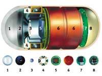
Capsule interior
- Optical dome
- Lens holder
- Lens
- Light recorder
- Battery
- Image recorder
- Transmitter
- Antenna
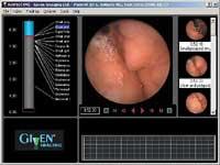
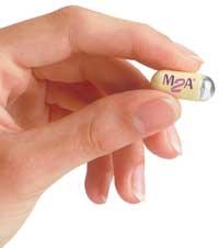
The most prominent example of the possibilities offered by the advancement of technology to carry out specific diagnoses can be a technique that is not yet used in conventional health services: the camera pill. It is another example of the topic in which reality surpasses fiction. In fact, in 1966, film director Richard Fleischer told the story of scientists who designed a microscopic size: that submarine entered the blood of the sick to find diseases and to be able to perform a first-hand treatment. The camera pill does the same at the base: the patient devours and takes pictures and the sample on a small screen complements the path of digestion. It is a simple pill, the size of conventional pills, that carries inside a microchamber that crosses the digestive system. In five hours the microcamera goes all the way and shows the images. This information allows the doctor to make a thorough diagnosis and subsequent treatment.
The microcamera was designed four years ago at the Royal Free Hospital in London. During this time he has carried out at least 20,000 explorations. Its size is 11 by 26 millimeters. This receives the signals and sends them to a wireless recorder. Most of the time it will be the patient himself who carries the recorder tied at the waist. The expert doctor will then use specific software to analyze the recorded images. On a five-hour journey, the pill records about 60,000 images, certainly more than can be obtained with any other system. Once the tour is completed, the pill leaves the corresponding path. The camera that is swallowed lets you see the entire small intestine. This represents a spectacular advance, since with the best techniques available so far only 30% of the small intestine could be seen. For some diseases, such as Crohn's disease or digestive bleeding, it is essential to see the small intestine well. The truth is that until now these diseases were perceived when they were very advanced, when much of the small intestine became patent. Therefore, treatment was needed after the production of the damage, while with the endoscopic pill diagnoses can be made in the initial phase of the disease.
In addition to the speed and accuracy of diagnostics, the new technique has more advantages: making painless observations, comfortable tests and not using radiation in observations. The advantage is that the tablet with microcameras presents difficulties, especially when it is said that the technique is very expensive, and the possibility of taking samples is not yet offered. Therefore, it is not yet used in most hospitals.
However, given the advancement of technology, it is clear that the failures of the new technique will be resolved at some point, that the technique itself will have more resources and that it is a good sample of future diagnostic systems.
Zu idazle
Zientzia aldizkaria