

Eye and camera of photos: are there similarities?
The camera, as an optical tool, is constituted by an optical system and a photosensitive receiver. The optical system (OBJECTIVE) is responsible for projecting on the receiver the real and inverted image of the object. The receiver, for its part, is formed by a plate or film covered by a layer of emulsion sensitive to light that will give us a reproduction of the object through a process of development.
The eyeball is also an optical or objective instrument. The coupling or coupling between the cornea and the crystalline forms a convergent or unifying system that generates a real and inverted image on the retina. As can be seen in this comparison, the retina or network acts on the human eye as a photosensitive receptor.
In this paper we will try to mention the different analogies between the eye and the camera, to later analyze the qualitative responses of an ametrope eye (abnormal eye) to different stimuli.
Basic theory
The optical system of the eye offers eight surfaces to the ray of light or to the visual stimulus, two corresponding to the cornea before reaching the net and six corresponding to the crystalline one. However, since the face before the cornea and the anterior and posterior faces of the lens are much more powerful than all other surfaces, we can say that to facilitate the separation between the cornea (which separates the air from the aqueous humour) and the crystalline (aqueous humore soaked from the anterior part and vitreous humore from the posterior part) it is necessary to differentiate it.

GLOBE OF EYES
Iris 1 Cornea 2 Front camera with aqueous humor 3' Crystalline (lens) 4 Sclerotic 5 Coroids 6 Retina (betsarea) 7 Phovea 8 Vitreous humor 9 Optic nerve
On the other hand, we must mention the retina or the grille: this structure is a photosensitive receptor, formed by visual cells, specifically the batons and cones. These visual cells are associated with bipolar cells and these in turn to nodal cells. As for the photograph of the retina, it must be said that the canes and cones are not distributed in the same way: while in the fobe (middle point of the eye, called yellow macula or blind spot) there are only cones, as we approach the periphery the number of canes increases.
The different distribution of these visual cells and the so-called duplicity of the retina, makes the phobe, in the center, concentrates the vision for large amounts of luminance or light, the detection of details and the vision in color (PHOTOPIC VISION) and the peripheral, on the other hand, is easier for the perception of low luminances, the vision in black and white (SCOTOPIC VISION).
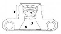
The camera, as can be seen in the image, can be functionally compared with the human eye in a relatively simple way. The lens of the camera (1) plays the cornea/crystalline coupling function; the diaphragm (2), its function is similar to that of the iris; the dark camera (3), equivalent to the sclerotic and the shutter (4), whose function is to regulate the exposure time. Finally, behind the shutter is the photographic film (5), the last receiver of optical information, as well as the retina in the eye. Following the analogy of the eye and camera, it can be said that the process of revealing to obtain the last photographic image is very similar to that of the nervous impulses that generate vision in the eye.
However, the retina or photosensitive receptor is the one that establishes the main differences between the eye and the camera. Unlike the photographic emulsion, due to the special distribution of the batons and batons and the special neuronal unions, not all regions of the retina have the same value when creating the image, while in the photographic system the answer is the same regardless of the part that participates. There are also photometric differences between the eye and the camera. While the effect of the light energy on the emulsion is aqumulatory, in the network the response of the visual cells is of instanpata, due to their ability to adapt these cells to high and low luminances.
Physiological processes
The eye, as a living optical instrument, has a coupling capacity or adaptation. And what is coodation? Ability to obtain sharp, accurate images of objects located in the visual field, in different places or points. The coupling process is accompanied by a series of physiological changes in the eye, such as the decrease of the beginini, the longitudinal displacement of the iris and the crystal, and in particular the variation of the capacity or power of the crystal. With all these changes the eye is able to accurately see all objects located between infinity and a nearby point.
The photographic system also has a coupling capacity. As in the eye, in the acoodation or focus mechanism, the diameter of the inlet globe decreases, with all the variations that it entails, the size of the mixing circle decreases, especially with the longitudinal displacement of the transparent media.
Let's finally see the problems of detail vision. Visual acuity is defined as the ability of the eye to detect and differentiate the smallest details of objects. The visual rigour of a person depends on many parameters, such as luminance, contrast between the test and background, size of the balloon, and also education of each person.
To distinguish two points close to each other it is necessary and necessary that at least one point not excited between the two visual cells excited by their images. In this sense, the state of adaptation of the eye and the retinal region used are of great importance, which contrasts with a camera.
In the photographic system, in order for the camera to appear as a separate or separate image of two points, it is necessary to intercalate between two impressed emulsion clusters, at least one unimpressed mole. As previously said, the camera cannot perform the same function as the eye in this sense.
Pathology Pathology
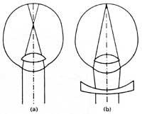
The eye emetrope (or normal) is one that is able to clearly see distant objects without performing any kind of appellate work. When this is not the case, we can say that we are in front of an ametrope eye (ametrope is abnormal). By specifying a little more, an eye can be near-sighted (with difficulty to see at a distance, since the object of the images is formed in front of the retina) or hypermetropea (in this case the close vision is affected, since the object of the images is formed behind the retina).
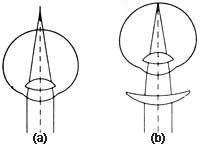
Another error is astigmatism. The characteristic of this ametropy is that the image of a point is not a point, but two, also located on two perpendicular lines. That is why those who have astigmatism do not see it clear.
(I want to thank my friends Joxe Mari Zinkunegi and Txaro Maiz for their collaboration in the elaboration of this article).
Zu idazle
Zientzia aldizkaria