

X-ray microscope
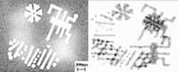
The current view of atoms is done through special electronic microscopes, but it is only possible under certain circumstances. And many have put in the X-rays the hope of seeing all atoms. The new microscope of researchers at Stanford University can meet these expectations. The device works with x-rays and visualizes the local structures of the materials. The first microscope of this type was developed in 1999 by the same group of researchers. The one at the time collected two-dimensional images with a resolution of 70 nanometers (nm).
The one presented in the journal Physical Review Letters, however, can observe structures ten times smaller in two dimensions, with a resolution of 50 nm in three. With this capacity the microscope is able to differentiate structures of 8 nm. While not yet seeing atoms, increasing the intensity of X-rays or lengthening the exposure time, researchers believe they will achieve the goal. The problem is that not all samples can be bombarded for a long time. Robust materials like semiconductors do, but cells do not. High-intensity X-ray lasers will be more suitable but in development.
Zu idazle
Zientzia aldizkaria