They have shown that studying the retina can be useful for monitoring parkinson's
Currently, in Parkinson's, the identification of patients at risk of cognitive impairment is a great challenge to provide more effective clinical treatments and advance clinical trials. To this end, researchers from the UPV/EHU and Biobizkaia have conducted a study based on the visual system, and have concluded that a method commonly used to perform ophthalmology tests may be adequate to monitor the neurodegeneration produced in Parkinson's. In fact, you've seen that retinal neurodegeneration can precede cognitive decline.
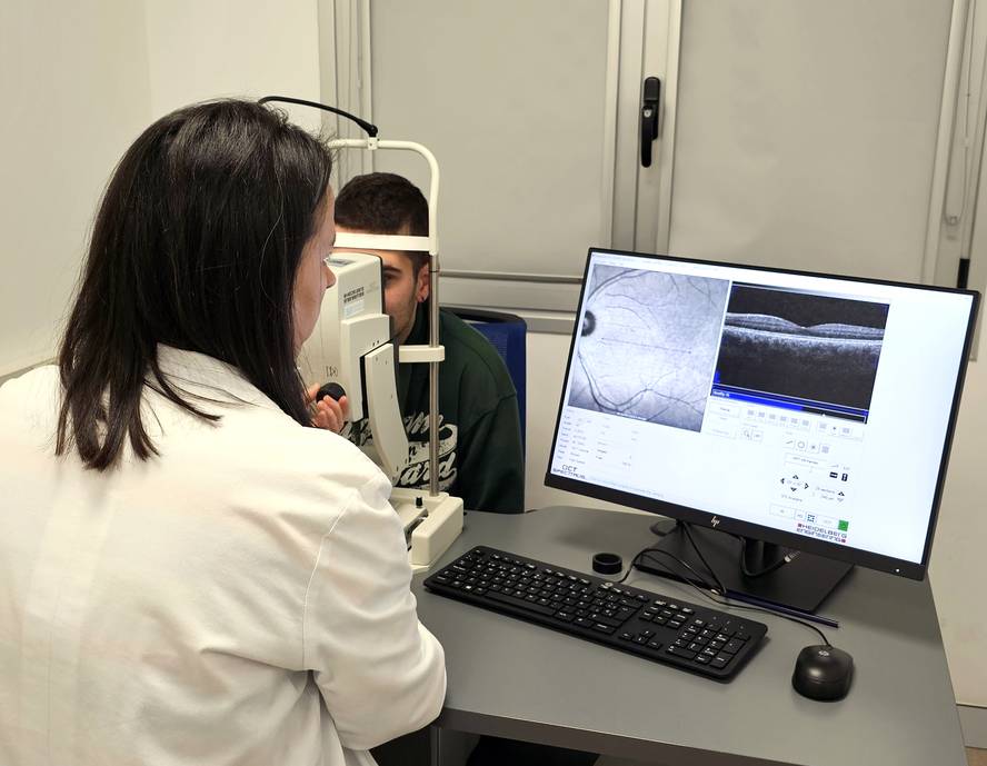
During the research, a group of Parkinsonian patients has measured the thickness of the inner layer of the retina using optical coherence tomography. This type of tomography is an instrument commonly used for ophthalmological tests and allows for repetitive and accurate high resolution measurements. Thus, in people with and without Parkinson's, the evolution of this retinal layer between 2015-2021 is analyzed. In addition, they have confirmed their results with images of the retinal layers of patients at a hospital in the UK.
The study shows that the retinal layer is remarkably thinner in patients with Parkinson's disease. In addition, it has been observed that in the early stages of the disease the major neurodegeneration is detected in the retina, and from a moment on, when the retinal layer is very refined, this neurodegeneration is performed as stabilized. Therefore, the slower refinement of the retinal layer indicates a greater severity of the disease.
Researchers have warned that to be used clinically it is necessary to confirm some aspects and improve resolution, but they have considered an important research, as the method they propose is non-invasive and present in all hospitals. Therefore, they continue to investigate to improve the method and to validate the results internationally.


