

Do you also draw symmetrical hearts?
From time to time, on a birthday letter or in the steam that the shower has left in the mirror, you've surely drawn a heart. Or that's what we've been told it's a heart.
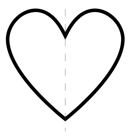
Observe carefully (Figure 1). You know, of course, that this is a simplification of the organ called the heart and that we actually use it to show more complex concepts than those that can be assigned to the organ. But have you ever thought beyond that? What are the characteristics that somehow turn this drawing into a round lie? From today you may draw simplified but at least asymmetrical hearts as a gesture of gratitude to your heart.
Left-right asymmetry of the human body
We tend to think that faces and symmetrical bodies are more beautiful, but asymmetry is a feature rooted in the human body in terms of internal anatomy. It is produced on two levels: in terms of general location and isolated organs. Take as an example the volume of the body as a whole and draw a left-right axis (Figure 2).

At this point it should be noted that in the medical language, the right and left are based from the point of view of the body being described. If we look at an individual face-to-face (as in Figure 2), the right of the individual is on our left and vice versa. Taking this into account, in Figure 2 we can see that the liver is located on the right side, stomach and spleen on the left side. For example, if you look at the internal organs alone, the lungs have three lobes on the right side and two on the left. The heart is asymmetric for both criteria. On the one hand, the highest volume in the left margin. In addition, there is inclination, that is, the left-right axis of the heart is not parallel to the vertical axis of the body (Figure 3). On the other hand, despite having four chambers, the right and the left chambers are different, both because of their histology and because of the traffic connections (arteries and veins). It should be taken into account, for example, that the tricuspid valve separates the right atrium from the right ventricle, i.e. it is composed of three segments, and the viculum (or mitral) between the left atrium and the ventricle is composed of two segments.

Physiological importance of cardiac asymmetry
Of course, attention to asymmetry goes beyond underlining the obvious characteristics, as it has great physiological importance. The asymmetric structure of the heart is necessary to establish what we call double circulation, and thus the organ performs its function: to spread the oxygenated blood to all parts of the body (Figure 4).
The left ventricle strongly pumps oxygenated blood to the aorta (number 1 in Figure 4), which will branch over and over to form smaller and capillary arteries that will reach all cells (number 2). When cells have taken oxygen, low blood in oxygen returns to the heart through the vena cava (number 3). This first cycle is called systemic circulation to the dispersion of oxygenated blood to the body and to the restitution of blood to the heart of “residues” with low oxygen content. Once the blood lowers in oxygen has reached the right atrium, it passes into the right ventricle through which it is pumped through the pulmonary artery (number 4) so that the blood will oxygenate in the lungs and return to the left atrium in the pulmonary veins (number 5). This second cycle, that is, the displacement to the lungs to restore oxygenation, is called pulmonary circulation.

Let us therefore return to the role in which asymmetry plays in ensuring that dual circulation is produced correctly. On the one hand, regarding the histological characteristics, it is necessary for the left ventricle to be more muscular, as the pumping force must provide enough pressure to the blood to reach the most remote cells of the body (and, on the contrary, the pumping force of the right ventricle must be only sufficient to reach the heart to the lung). On the other hand, the aforementioned chamber artery, as explained in all chamber waiting connections, should only be produced in this way, so that the circulation is adequate.
Genesis of asymmetry in embryonic development
Taking as a reference the three axes that define the three-dimensional space, the left-right axis is the last defined in embryonic development. An embryo is at first a perfect sphere in which cells are placed homogeneously. It's growing, doubling the cells, but it's just a mass without known order. If there is no asymmetry, no axis can be defined or, rather, the axes can be defined in any way. Throughout development, some molecular signals allow the emergence of characteristic structures in the embryo that define the inferior upper axis and the forward axis. Among others, the creation of the primitive line establishes this structure [1] (Figure 5).

When two axes have been defined, the third axis is established with force, since there is only one way to establish a third line perpendicular to two straight lines in space. At that time, the left and right parts of the body are defined, but at the moment, they're completely symmetrical. In the third week of human embryo development, precursor cells of the heart begin to form a cardiac tube in the centre of the left and right axis of the embryo [2], [3]. This pipe is located at the beginning in a straight and vertical way. Then, it starts to fold up to the loop shape (Figure 6). This fact is of great importance because (1) it establishes a precursor form of the heart (morphology), which is the first functional organ that occurs in the body, and (3) is the first asymmetry of the left to the right seen in the embryo.

As the cardiac loop develops, a mature heart is finally produced with the exact anatomy described above. In addition, all the asymmetric organs mentioned above will create their own form and develop their own asymmetric characteristics. All these processes are the object of study in embryonic - morphogenic science and are important not only because they provide us with basic information about our origin, but also because they help us understand the causes of wrong development.
Developmental defects and pathological symptoms
What has been described so far is an explanation of what happens in healthy humans. In the medical language we use the Latin expression situs solitus to refer to the distribution of organs of a human being that has developed regularly (Figure 7). Defects that alter left to right asymmetry may appear in embryonic development. The average frequency is 1 case per 2000 inhabitants [2]. These disorders may have a systemic impact, i. e. may cause malformations in all asymmetrical organs of the body, or in some cases. If a total displacement occurs, that is, if an individual has left and right displaced, we describe it as a mirror image with the expression situs inversus totalis. If some organs do and others do not, they move in a confusing and unconcordant way, we will find a state of situs ambiguus or heterotaxis [4].

We have described in general the possible pathologies of the internal organs, but the same principle can be applied specifically to the case of the heart. If an individual presents situs inversus totalis, by definition, all organs, including the heart, will be displaced. If the mirror image is perfect and, therefore, all arteries and veins are connected to the corresponding chamber, the function of the heart is generally normal. The case series of heterotaxia, on the contrary, is very extensive, so the prognosis is very varied [5]. Sometimes, we can find severe heart malformations. They are called congenital cardiopathies and have their origin, at least in part, in inadequate forms of the cardiac loop. To cite two examples, the exchange of position of the aorta and pulmonary artery, or the fact that the muscle wall separating the left and right parts of the heart is not completely closed and has “holes” (Figure 8). Fortunately, some malformations have been resolved surgically, while others are excessively complex, so the patient will have to live with this defect and often lead to premature death.

What can science do for all of this?
A better understanding of the causes of congenital heart disease is essential to be able to make a faster diagnosis in newborns and to offer more effective surgical treatments. Medicine has come a long way to this day, but there are still many cases that cannot be solved.
To deepen this knowledge, as is usual in the study of biosciences, animal models are used. In the heart development study, researchers study animal models with genetic variations (mutations) and measure the specific excitation that each variant produces in development. These resources allow us to know more and more precisely this process of converting the heart loop into a mature heart. In addition, the approval of the animal and human genes allows predicting the effect of human mutations on development, which may allow early diagnosis at some point. Research, however, requires considerable investment in time and money. In return, we have a good journey to understand the retribution for the development of medical advances and the complexity of biology. If beauty is inside, we can say that beauty is asymmetrical.
Bibliography
1] Shahbazi MN. Mechanisms of human embryo development: from cell fate to tissue shape and back. (2020) Development. 147(14):dev190629.
[2] Bleeding A, et al. (2018) Left-right asymmetry in heart development and disease: forming the right loop. Development.145(22):dev162776.
[3] Bleeding A, et al. (2020) Transient Nodal Signaling in Left Precursors Coordinates Opposed Asymmetries Shaping the Heart Loop. Dev Cell. 55(4):413-431.e6
[4] Djenoune L, et al. (2022) A change of heart: new roles for cilia in cardiac development and disease. Nat Rev Cardiol. 19(4):211-227.
[5] Lin AE, et al. (2014) Laterality defects in the national birth defects ion study (1998-2007): birth prevalence and descriptive epidemiology. Am J Med Genet A 164A(10):2581-91.
Zu idazle
Zientzia aldizkaria