Organelles without membrane
When cell biology schools explain cells and their characteristics, two main types of cells are always mentioned: eukaryotic cell and prokaryotic cell. And on the other hand, when the differences between both types of cells are listed, organelles appear before or after. Organelles are often defined as specialized structures that perform specific functions inside the cell. Explanations of organelles have always mentioned the nucleus, protector of cellular genetic information; mitochondria and chloroplasts, energy producers; the granular and smooth endoplasmic reticulum, involved in the synthesis of lipids and proteins; and the Golgi apparatus, composed of secretion vesicles that form and distribute protein packages. They all have a specific function and are surrounded by a semi-permeable membrane.
In the last 100 years there have been no changes in the cellular structure we know. Or yes? In November 2018, researchers at the Sloan Kettering Institute published a new organ in the journal Cell (Ma & Mayr, 2018). New organelle assigned to a TIGER domain (which we will explain later)! Would we be facing a new concept that would change the integrity of the cell?
In recent years, researchers have looked more closely at the water soup and molecules of both cell cytoplasm and organelles. Researchers have observed a large number of solid vesicles in the cytoplasm and inside the organelles (i.e., inside the organelles with a certain density surrounded by liquids of another density), which, like membranous organelles, have specialized functions. These vesicles often do not correspond to the definition of a “classic” organelle: on the one hand, these non-membranous organelles have no concrete limits, and on the other, they can be ephemeral organelles, they can react quickly under the influence of chemical signals and, at the same time, disorganize specialized structures generated in the disappearance of the stimulus.
However, because of their function and importance they have achieved the attention of researchers.
There are several organelles without membrane
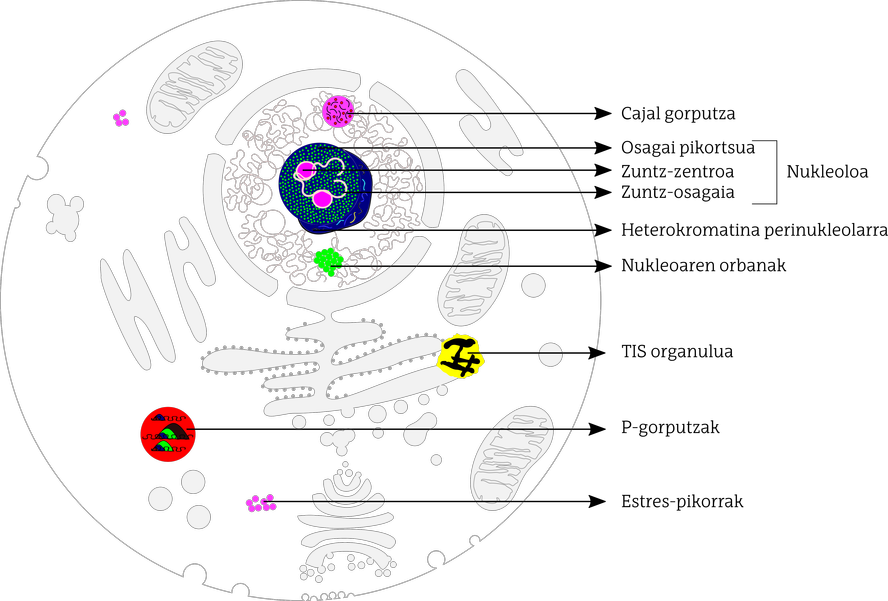
In mammalian cells, researchers have identified at least a dozen membrane-free organelles dispersed in both the cell nucleus and cytoplasm (Figure 1). In general, non-membranous organelles within the nucleus are specialized in controlling gene transcription and in some aspects of RNA metabolism. However, non-membranous organelles dispersed by cytoplasm intervene in the metabolism, transport and homeostasis of messenger RNA.
a) Membrane-free organelles within the nucleus
Among the non-membranous organelles within the nucleus, the nucleolus stands out. The nucleolus is the most studied and probably the largest of all. Its main function is the biogenesis of the ribosomes needed to synthesize proteins. In the nucleolus three regions are distinguished: fiber center, dense fibrous component and granular component. In the first, the DNA encoding ribosomes is located; the conversion to RNA of this DNA, the transcription, occurs at the boundary between the fiber center and the fiber component. In this limit appears the protein known as fibrilarine and RNA processing is produced that encodes ribosomes, adhesion to the cut. The granular component, on the other hand, is rich in nucleophosphate proteins, where the accumulation and assembly of pre-ribosomal subunits occurs. The nucleolus is surrounded by very compact organized DNA, called perinucleolar heterocromatine. They are mainly rich in DNA repeats, mainly DNA satellites and ribosomal DNA sets. It seems that this repetition of ribosome DNA prevents the conversion or recombination of DNA, stabilizing the ribosomal DNA.
Cajal bodies are also found in the nucleus of the cell, but they are smaller. As for the structure, they consist of proteins and RNA that appear to be necessary for the assembly and modification of snRNP (small nuclear ribonucleoprotein). These snrnp are necessary for post-transcription changes of messenger RNA: among others, transcribed parts of RNA that will not become proteins, introns, cutting adhesions. Cajal bodies appear to participate in the final maturation process of snRNP particles and in the generation of snRNP complexes.
Nucleus stains are directly related to Cajal bodies and are part of the nucleoplasma. In the stains of the nucleus accumulate and modify snRNP and proteins rich in amino acids serine and arginine. They all participate in the cutting and gluing process of the messenger RNA.
b) Membrane-free organelles dispersed in cytoplasm
As for non-membranous organelles dispersed by the cytoplasm, they are usually called mRNP granules (messenger ribonucleoprotein). Although there are several types of RNP, it is common for proteins and messenger RNA to be shared and interacted.
P bodies (processing bodies) have been described in different cell types and have been observed to be protein-rich areas involved in the transport, modification and translation of messenger RNA. When many untranslated messengers to amino acids accumulate, either because the translation is inhibited or under certain stressful conditions, the size and number of P bodies increase.
Stress granules, as the name suggests, are assembled before stress signals, sequestering muted messenger RNA molecules and translation factors. That is, the cell stores in specific areas the material needed to cope with this stress. In stress granules it is usual to find the necessary factors to start translation and components of small subunits of ribosomes.
Other membrane-free cytoplasmic organelles are germ granules, which appear in the germ cells of the developing embryo (sex cell stem cells). They are usually rich in messenger RNA and enzymes that modify RNA. It seems that they intervene in subsequent changes to the translation of messenger RNA into germline cells.
In September 2018 they published in Cell magazine the discovery of the latest type of membrane-free organelles: It was known as the TIS organelle. In interaction with the endoplasmic reticulum, it allows interactions between proteins directed by the 3’UTR end of the RNA. TIS11B protein is closely related to this organelle, hence its name. The interaction between the TIS organelle and the reticulum is called the TIGER domain. The latter is different from other cytoplasm media, both biophysical and biochemical. All this will control the exchange of proteins of the endoplasmic reticulum, regulating the interactions between proteins, relating the 3’UTR extremes with the protein TIS11B and organizing a network in the cytoplasm and around the smooth reticulum.

Related to diseases
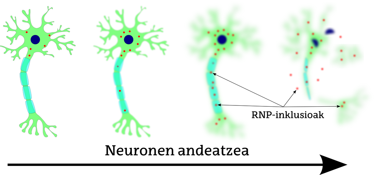
The high dynamism of non-membranous organelles makes it not easy to establish direct relationships between functional problems and diseases of these organelles. However, the case of amyotrophic lateral sclerosis is noteworthy. One of the characteristics of this disease (such as other neurodegenerative diseases) is that inclusions of ribonucleoprotein messengers (RNP) occur that alter the normal metabolism of messenger RNA (Figure 2). These pathological inclusions are formed by RNP protein granules, whose large accumulation produces an assembly between them and the formation of amyloid fibers. Amyloid fibers alter the normal functioning of cells, degrading them.
In cancer cases, non-membranous organelles also present interesting options or perspectives. One of the factors that govern cell growth is the assembly of ribosomes, which as has been seen has a great importance in the production of ribosomes. The nucleolus, therefore, can be an important destination for cancer treatments, characterized by the uncontrolled growth of the nucleolus and the increase of the nucleolus.
The key to the birth of life? Rethinking Oparin
In 1922, Russian biochemist Alexander Oparin published a theory on the origin of life at the beginning of Earth's history. According to his theory, the first simple organic compounds were formed, since the electric energy of the rays or the heat energy of the volcanoes provoked the reaction of methane, ammonium, water and other components, rich in the reducing atmosphere of the time. All these ingredients were arranged in small drops called coacerbatos. In 2016 a group of German researchers described the chemically active and liquid droplets with fragmentation capacity and therefore perpetuation. These drops and non-membranous organelles have some equal characteristics, so will there be keys to explain the origin of life in membrane-free organelles?
References
Crabtree, M., Nott, T. 2018. These organelles have no membrane. The Scientist. December 2018. https://www.the-scientist.com/infographics/infographic--what-are-membraneless-organelles--65135
Gomez, E., Shorter, J. 2018. "The molecular language of membraneless organelles". Journal of Biological Chemistry 1-23.
Ma, W., Mayr, C. A. 2018. "Membraneless Organelle Associated with the Endoplasmic Reticulum Enable 3< UTR-Mediated Protein-Protein Interactions". Cell 175: 1492-1506.
Mao, Y.S., Zhang, B., Spector, D.L. 2011. 'Biogenesis and Function of Nuclear Bodies'. Trends in Genetics 27: 295-306.
Mitrea, D.M., Kriwacki, R.W. 2016 "Phase separation in biology; functional organization of a higher order". Cell Communication and Signalling 14: 1.


