

New microscopy technique to see all cells in an area of the brain
The scientific journal Cell reports a new microscopy technique that allows to see for the first time all cells in an area of the brain. It is the result of a study carried out by the UPV and the Basque Achucarro neuroscience research centre in collaboration with the University of Bordeaux.
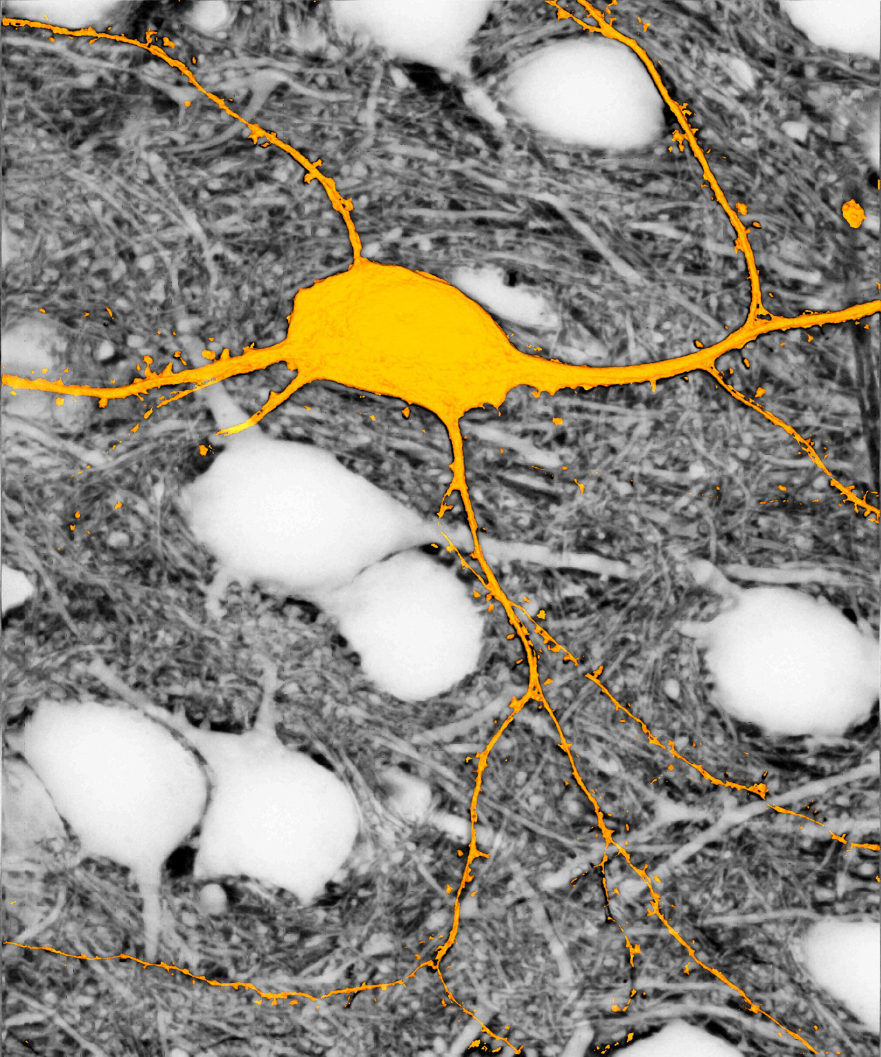
When investigating the living tissues of the brain, microscopy methods so far only allowed to see previously marked cells. The technical limitations prevented labeling all cells in a certain region of the brain, which greatly conditioned the vision of the brain: one could not see how the cells of the brain are organized and how they act. Many spaces appear blank in the images of microscopy.
Now, with the new SUSHI microscopy technique (acronym for “Super-resolution Shadow Imaging”), scientists can easily label the space surrounding brain cells, loaded with fluids, without having to be marking each of them. In this way, all the cells of the brain zone located in the microscope and their interactions can be seen. This is especially interesting, since the extracellular area of the brain presents a particularly complex structure and although its function is physiologically important, its structure and dynamics has so far been very difficult to analyze.
Zu idazle
Zientzia aldizkaria
- Babesleak
-

-

Elhuyar
Nor gara | Kontaktua |
Publizitatea
| Laguntza
Pribatutasun politika | Cookien politika
ISSN-2603-6614 Elhuyar