

New imaging techniques for the investigation of respiratory diseases
The lungs are a basic organ for animals that breathe air, including humans. Lung diseases usually have a great influence on the whole body, as they hinder the exchange of oxygen that cells need to obtain energy and carbon dioxide, residue of breathing.
In rich countries, chronic obstructive pulmonary disease (from chronic buxctive pulmonary disease, COPD) is the lung disease that produces more deaths, apart from infections. This disease limits the flow of air and is related to chronic bronchitis and emphysema. Bronchitis is the inflammation of the respiratory tract and emphysema causes the degradation of lung tissues. On the other hand, in the poorest countries, lung infections are the main cause of death, above any other disease. In addition, the world's deadliest cancer is the lung.
Like any other disease, there are several possibilities of research on this subject. On the one hand, it can be done at cellular level. The latest developments in genetics, proteomics, etc. They allow to study the molecular and microscopic components of the disease. On the other hand, clinical studies can also be carried out, for example, applying different treatments to different groups of patients and analyzing the results. However, both methods have their limitations. Although cell research can be highly accurate, it does not explain the interaction between cells and tissues throughout the body. And studies carried out in the clinical phase pose tangible ethical problems, such as the non-administration of a drug that can benefit a group of patients or the demonstration of potentially harmful therapies. To fill the gap between both methods, animal trials are usually performed. Thus, from 1901 to 2006, 76% of the Nobel Prize winners of Medicine used animals in their research.
The use of mice, rats, etc. Before going to the clinical phase it facilitates many tests, but of course, the ethical problems do not disappear at all. For this reason, in English the so-called "principle of three R" is applied: reduce as much as possible the use of animals ( reduce ), replace it with other techniques that can give the same information ( replace ) and refine the treatment given to animals ( US$ ).
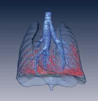
In this context, new imaging techniques have a lot to say. High-resolution magnetic resonance techniques or x-ray based scanner (called micro-CT) allow, among other things, to analyze the body of small animals without causing pain in them, which is what is called refining the treatment. In addition, by not having to kill animals in the experiments, the number of animals used can be significantly reduced. The microct is especially suitable for the study of small animal lungs, with a resolution of approximately 50 m (0.05 mm), in addition to showing a great contrast between lung tissues and air.
Drawing technique or how to obtain a pulmonary image of a mouse
High-definition micro-cts have developed mainly in recent decades, but their main principle is the XX. Known since the early twentieth century. In the first step, several X-rays are made of the object to be studied, forming a circumference from several angles and around the object. Subsequently, these x-rays must be processed by computer to obtain a three-dimensional image of the internal structure of the object. The more X-rays you make in the first step, the more straight the final image will be.
To access the scanner, animals are usually anesthetized, but breathing movements can cause problems, especially in lung images. In fact, if on each X-ray the lungs are in a different position, the three-dimensional image processed by the computer will have little precision, since somehow it will show approximate lungs. To avoid this we have developed a technique of respiratory synchronization and image that consists of connecting the animal to a respirator once anesthetized. To do this, a catheter is inserted into the mouse trachea, which is narrower than a millimeter. To facilitate the operation, we use fiber optic illumination inside the catheter.
When the animal is connected to the respirator, the scanner can be accessed. To prevent the lung movement from affecting the image, it is necessary to synchronize the respirator and scanner. By means of an electrical signal, x-rays are obtained only at the time the lungs are filled. Thus, in all x-rays taken from different angles the lungs are in the same state and the three-dimensional image calculated on the computer will have a much better resolution.
Imaging techniques or how to measure the incidence of a disease in the lung image of a mouse
By observing the images of the lungs of animals we can obtain important information. But in an experiment, differences between two or more groups are usually sought: for example, if a treatment has slowed or, at least, has slowed the development of a disease. In this type of research it is very useful to have quantitative data and objectives that allow an equitable comparison. Therefore, it is very important to obtain objective measures from images. For this we have used the diagnosis by computer. We have developed several algorithms to obtain, step by step, measures that indicate the state of the lungs from the initial image.
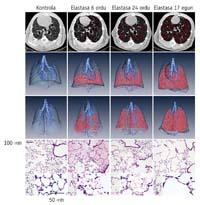
Before making a measurement, we must separate the sections that interest us through a process called segmentation. In this case, it is advisable to segment in the figure the lungs and the airways in order to make measurements on them. In the case of the airways, we have developed an algorithm based on field growth. The computer program is able to automatically locate the trachea and from there it goes following the airways to detect the entire bronchial tree. We have developed a new program of separation of lungs that seeks in several steps a dark zone in the center of the image. To evaluate these automatic techniques, we compare them with manual segmentation.
When, in the image, the airways and lungs are separated and identified, measurements can be made. The airways allow automatic measurement of bronchial diameters. In the lungs the most interesting measurements are volume and intensity, since the intensity of the micro-CT images (the luminosity of each pixel) is related to the air content. The darker, the more air. Thus, the inflamed lungs usually have a high intensity and those of low emphysema, since the degradation of the tissues characteristic of the disease produces a greater proportion of air.
Experiments or how to apply these new techniques to real experiments
The techniques developed to obtain precise images of the lungs of the mice and obtain objective measures of the mice have a practical application, since in addition to reducing the use and treatment of animals in the experiments of study of diseases, they offer a rich information. To demonstrate this, we conducted different experiments, especially with two diseases: chronic lung inflammation and emphysema.
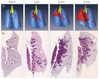
There are various techniques to cause chronic lung inflammation in the mouse. One of them is related to silicosis that occurs in humans and consists of introducing silica crystals in the lungs of the animal. This causes a chronic inflammatory process of several phases. For the first time around the world, we have used micro-CT technique and automatic image analysis to describe this disease pattern, obtaining new development data. In summary, we found that inflammation narrows the airways and that the proportion of inflammation of the lung increases for several months.
In the case of emphysema, the disease occurred through an elastase that disappears the lung tissues. In previous studies, several researchers showed that the micro-CT technique could be used to study emphysema development, but it was not practically compared with other techniques. We have carried out extensive research using different techniques at the same time, and have found that micro-cts shows with great precision the development of the disease without the death of animals.
Conclusion or what we have learned with all this
Lung diseases cause many deaths worldwide. In our work we have developed techniques to be able to investigate them better using small rodents. On the one hand, we have developed a method for obtaining high precision pulmonary imaging using X-rays, synchronizing artificial respiration with x-rays. On the other hand, we have developed automatic computer programs for the analysis of these images, in order to obtain objective measures. The usefulness of these techniques has been demonstrated in different experiments.
Accelerating experiments with animals, refining and obtaining richer information, these techniques will facilitate the passage of the healing treatments of lung diseases from the laboratory to the clinical phase.
BIBLIOGRAPHY BIBLIOGRAPHY
Zu idazle
Zientzia aldizkaria