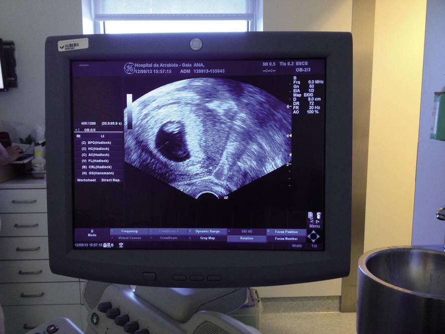
1% fetal loss dogma, decline
The quarterly journal Prenatal Prespectives of the International Society for Prenatal Diagnosis (ISPD) published in 2014 the following article: Invasiveness: the decline of the dogma of the 1% Fetal Loss Rate 1% fetal loss dogma, decline).
The authors of this article, Borrell and Stergioutou, have analyzed several published papers on invasive techniques, amniocentesis and risks of fetal loss attributable to the corial biopsy, reaching the following conclusion: The risk of loss attributable to these techniques is 0.1% (1/1,000), with no significant differences between the fetal loss of pregnant women to whom invasive methods are applied and those who are not.
They have also concluded that it is critical to provide women with accurate and up-to-date information on invasive and non-invasive techniques that enable them to make evidence-based decisions. It is said that fetal loss values that are not real for invasive techniques should be excluded from clinical activity, as well as misleading diagnostic capabilities for non-invasive ones.
It has also been proposed to prioritize the appropriate training of specialists on invasive methods. In this way, all prenatal diagnostic techniques can offer individualized selection and excellent results.
The authors recall that the increased risk of fetal loss from invasive tests is 1% and the origin of the “dogma”, long approved: An article published in 1986 by Tal, comparing the percentage of fetal loss of 4,606 women who suffered amniocentesis (1.7%) with that of a control group that did not apply invasive technique (0.7%).
Other articles, such as the one published by Eddleman in 2006, comparing the fetal loss of 3,096 pregnant women who were given amniocentesis before the 24th week of pregnancy (1%) and 31,907 pregnant women who did not perform this invasive test (0.94%). The conclusion was as follows: The increased risk of fetal amniocentesis loss was 0.06%.
For its part, a group of the University of Washington, in 2008, published results similar to those of Eddleman and concluded that there were no statistically significant differences between the fetal loss corresponding to the group to which the amniocentesis was performed and that corresponding to the control group (Odibo et al, 2008; Odibo et al, 2008b).
Finally, in 2011, the Nicolaides group (Akoldakar et al, 2011) showed that most of the fetal losses attributable to the chorial biopsy were predictable depending on the characteristics of the mother and/or pregnancy.
Based on these characteristics, a forecasting model of 33,856 pregnancies was developed between the number of expected fetal losses among women who had a comparative korial biopsy and those who did not apply this invasive technique. The study showed that there were no large differences between expected and actual amounts.
Likewise, this same group (Akoldakar et al, 2014) has just published a meta-study after a thorough review of various publications. The findings of this research have shown that there are no significant differences in fetal losses before the 24th week of pregnancy, among women who suffered amniocentesis (42,716 women) and biopsy (8,899) and those who did not suffer amniocentesis (138,657 women) or korial biopsy (37,338). According to the study estimates, in the case of amniocentesis, the fetal loss attributable to invasive techniques was 0.11%, while in the case of biopsies it was 0.22%.
Comments and conclusions
The difference between the first article (1986) and the rest (last of 2014) lies in the greater experience of professionals and in the evolution of the resources used, in the thickness of the needles and in a much greater resolution of the ultrasound.
Thus, the Committee Opinion of ACGO (American College of Gynecologie and Obstetricians) recommends the use of the array-CGH technique in pregnant women with ultrasound alterations that make an invasive prenatal diagnosis.
In 2014, Wapner reviewed the number of submicroscopic alterations detected in amniotic fluid using the array-CGH technique, with the following results: 0.9% on average in 12,000 low-risk pregnancies. Currently, the risk of aneuploidia 1/270 is assumed as the basis for an invasive prenatal diagnosis, based on a certain balance between genetic risk and the risk of fetal loss by invasive procedure.
Therefore, it seems appropriate to offer an invasive and array-CGH technique to all pregnant women. This attitude would share the recommendation of the American Congress of Gynecology and Obstetrics 2007, which suggests offering all women the possibility of performing an invasive pre-natal test without taking into account the risk.
The experience of the genetics unit of Policlínica Gipuzkoa (1,300 prenatal diagnoses by array-CGH technique in low-risk pregnancies) confirms the published results. We diagnosed nine submicroscopic pathological alterations (0.7%) that will not be detected if the array-CGH technique had not been used before birth.


