

3D virtual map of human embryo development
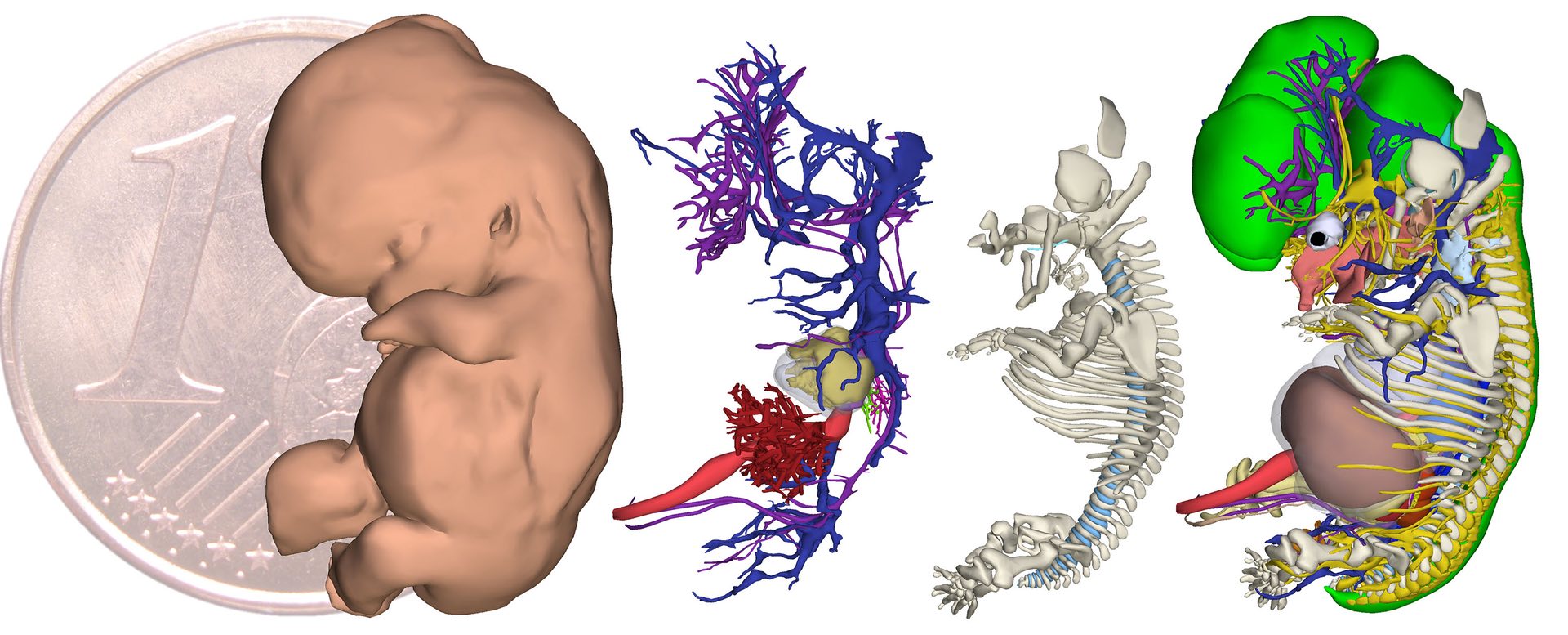
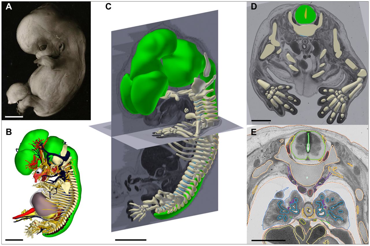
After analyzing the collection of 15,000 embryo samples, a group of researchers from the University of Amsterdam has created an interactive database of human embryo development. This 3-dimensional digital atlas accurately gathers the development of the first two months of human embryos, structure by organ and structure, about 150. You can see the project here.
In the creation of the atlas, questions have been clarified that until now were confused, such as the kidneys and the development of gonads. The researchers have stated that the Atlas will also serve to carry out research related to human development and its diseases.
Using this model, researchers have been able to verify that there are significant differences between human development and the embryo of mice and chickens. Taking into account that these are the models that so far have been used to analyze the development of mammals, they have given importance to the identification of these differences.Their work has been published in the journal Science.
Zu idazle
Zientzia aldizkaria